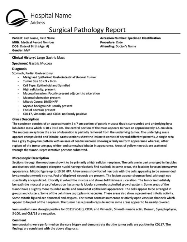
Sample of Pathology Report
After the diagnosis of GIST, a patient should take important steps to learn more about their particular disease so that they may find optimal care and treatment. This can be a very challenging and sometimes overwhelming task. Getting a copy of the surgical pathology report is one of those steps.
This report is generated after a surgery to remove GIST tumors and contains crucial information regarding diagnosis and key factors for calculating risk of recurrence. Understanding your risk of recurrence, or the chance that a tumor will return after surgery, is especially important when considering preventative or (adjuvant) Gleevec.
Pathology reports are written by pathologists (doctors who study the cause and effects of diseases) and identify the diagnosis based on their examination of the tissue sample from a surgery. The majority of pathology reports begin with a similar setup – hospital information on top followed by the patient’s information. An important part to notice in this section is the accession number. This is a specimen identification number that is unique for every patient and procedure. Pathologists use this unique number to identify tissue samples while performing tests.
Most pathology departments include the same major sections; however, they may be arranged in a different order. The following are examples of the most common sections found in a pathology report:
Diagnosis
Diagnosis is the summary of everything found during the pathologist’s examination of the tissue, including diagnosis details and tumor features (surgical margins, size, malignant potential, etc.). If there were several excisions made during the surgery (several tumors removed), there will be multiple entries under the diagnosis description for each one. This is a good place to look for an overall summary of the pathology report.
Gross Description
The gross description describes the tissue sample’s physical description when the pathologist receives it in the laboratory from surgery. This section may contain many medical words, however the key parts to look for are the size of the tumor and the tumor location. These factors are used when calculating your risk of recurrence.
Microscopic Description
The microscopic description is what is seen when the pathologist looks at the tissue under the microscope. This includes the types of cells and their condition (i.e. hemorrhagic). An important part in this section is the mitotic rate, or the measurement of cellular proliferation or cell division. This number helps determine how fast a tumor is growing and is one of the most import factors to consider when calculating risk of recurrence.
An additional test that may be performed is immunohistochemistry (IHC). IHC is the process of using stains to detect the presence or lack of particular proteins. For GIST, the most common IHC stains are C-Kit (CD117), CD34, and DOG1. Positive results for these proteins indicates the diagnosis of GIST. These results may be on a separate report, but are still a major factor in diagnosis.
Comment
Pathologists may include information for your treating physician. This will either clarify unclear results or recommend further testing to be done.
Clinical Information
Your treating physician may include clinical history that is relevant to the tissue that the pathologist is examining. This may include diagnosis, the nature of the disease, or other diseases that should be of concern.
Specimen/Tissues
This section indicates what was removed during the surgery and where it was located. For example, a tumor removed from the stomach may appear as “gastric tumor.”
A patient’s risk of recurrence, or the chance that a tumor will return after surgery, can be determined using several indicators (mitotic rate, primary tumor size and location). One of the key pieces of information is the mitotic rate. The higher the number, the quicker the cells are dividing, leading to faster tumor growth.
There are several different nomograms (tools for determining risk of recurrence) that can be used. Some nomograms consider other factors such as tumor rupture, surgical margins, and mutation. It is important to find the best nomogram to use based on the information provided on the pathology report. Based off of the Modified NIH Method, which is one nomogram to calculate risk, mitotic rates less than 5/50 HPF are considered low risk and anything greater than 10/50 HPF is considered high risk. However, since risk of recurrence is based off of multiple factors, conclusions should not be made without all the necessary information.
There are certain situations where mitotic rate is irrelevant and should not be taken into consideration when calculating risk of recurrence. One situation is when there has been metastasis or a recurrence already. This is because there is no need to determine a risk of recurrence when there was one already. The same way you wouldn’t check the odds of winning the lottery when it has already been won. Another situation is when a patient has received Gleevec or any other form of chemotherapy prior to having surgery on their primary tumor. This is because chemotherapies alter cellular division and tumor growth. If mitotic rate is determined after chemotherapy, it would provide a non-representative rate, often times lower than the actual. Mitotic rates are best determined from single primary tumors that have never been exposed to chemotherapy.
At first glance, a pathology report may seem overwhelming. However, once you know what to look for it becomes easier to interpret. After every surgery, always ask for a copy of the pathology report so that you may take the time to read through and discuss your risk assessment with a physician.


